In recent years, cell therapy has emerged as a transformative treatment, providing promising cures and even more potential for treating various cancers and other diseases. However, amidst the optimism, a significant concern looms—the presence of foreign particles in cell therapy. These tiny intruders, often difficult to detect, can pose serious threats to the safety of cell-based treatments.
These particles originate from many sources, such as contaminants introduced during manufacturing, aggregated proteins, or magnetic beads used to expand and activate cells. The consequences of their presence can be severe and multifaceted. Subvisible particles, measuring ~10 µm in size, have the potential to cause capillary occlusion or trigger unwanted immune responses in patients, resulting in treatment rejection, reduced efficacy, or, in worst-case scenarios, death.
Adding to the complexity is the fact that most traditional analytical techniques, such as flow imaging (FI) and light obscuration (LO), categorize cells and cell debris alike as particles. This leads to the challenge of having to find unwanted particles within a sea of desired ones.
Current regulatory guidance regarding particles in cell therapies has been ambiguous. However, the FDA’s 2023 warning letter to Novartis concerning particles found in its Kymriah® product is a clear indication that overlooking these hazards is no longer an option. In the letter, the FDA specifically highlighted particles from cryo bags, emphasizing that it is crucial to ensure these bags are completely free of particulate matter.1

Manufacturers urgently need robust solutions capable of fully characterizing subvisible particles throughout the development process. This ensures that therapies are safe and effective before being administered to patients.
Develop Faster, Smarter, and Safer with Innovative Particle Analysis and Detection
The good news is that recent advancements have led to the development of new particle detection methods that can easily and quickly distinguish particles in cell therapies.
For example, Halo Labs’ powerful imaging trio can uncover crucial subvisible particle data, leading to more accurate results and better insight:
- Backgrounded Membrane Imaging (BMI) brings clarity to formulation risks that were previously hard to see, breaking through traditional limits quickly. One minute is all it takes to analyze a sample, with the ability to test 96 samples in less than two hours.
- Fluorescence Membrane Microscopy (FMM), when combined with BMI, offers an unmatched level of analysis for identifying, counting, and sizing particles. FMM uses specific fluorescent dyes or antibodies to label aggregates, integrating seamlessly into existing workflows.
- Side Illumination Membrane Imaging (SIMI) is included with each BMI measurement, allowing for the characterization of every particle in your sample.
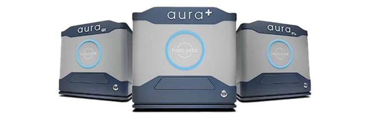
All of these technologies are exclusively available on Halo Labs’ cutting-edge Aura® particle analysis systems. With just 5 µL, subvisible particle analysis is possible at the very beginning of your process. This helps speed up development and allows researchers to make better, clearer decisions, preventing problems before they become expensive bottlenecks.
Aura CL Allows You to See More with Less
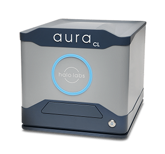
Measuring particles in cell therapy manufacturing has long been a challenge, and the fact that the drug product itself is a particle makes it even harder. Unlike traditional methods that can’t tell cells apart from protein aggregates or harm-causing particles, Halo’s Aura CL® is the first system that can identify them quickly while providing all required cytometric capabilities—counting cells, identifying types, and measuring viability in one simple assay.
With Aura CL you can:
- Detect and count residual Dynabeads. Traditional methods of detecting and counting residual Dynabeads can be subjective, labor-intensive, inaccurate, and prone to errors. Aura CL simplifies this process by providing high-speed accurate counts, enhancing your efficiency and accuracy.
- ID cells vs. non-cells. Manual sorting through images or mastering intricate machine-learning libraries can be time-consuming. Aura CL simplifies this task by precisely distinguishing cells from non-cell aggregates right from the start.
- Analyze small volumes. Aura CL only requires 5 µL of sample for analysis. This small volume requirement facilitates CAR-T characterization at an earlier stage in cell therapy development, saving valuable resources and time.
Conclusion
While the potential of cell therapy is undoubtedly groundbreaking, the dangers posed by foreign particles should not be underestimated. A comprehensive understanding of the risks and attention to quality control during the manufacturing process while utilizing the latest available analytical methods to detect particles is imperative to unlocking the true power of cell-based treatments. As we navigate new therapeutic modalities, acknowledging and addressing these challenges will be pivotal in realizing the transformative benefits of cell therapy while ensuring the safety and well-being of patients.
References
- https://www.fda.gov/media/174213/download
- https://bioprocessintl.com/bioprocess-insider/regulations/kymriah-gmp-issues-in-nj-led-to-fda-letter-for-novartis/
- https://www.fiercepharma.com/pharma/novartis-takes-fda-flak-after-inspection-turns-significant-kymriah-manufacturing-flubs
All product names, logos, brands, trademarks, and registered trademarks are the property of their respective owners. All company, product and service names used in this website are for identification purposes only. Use of these names, trademarks, and brands does not imply endorsement. Dynabeads refers to the magnetic beads produced by Thermo Fisher Scientific, Inc. Halo Labs is not affiliated with Thermo Fisher Scientific, Inc., and references to Dynabeads or any other third-party trademark do not imply sponsorship, endorsement, or approval.
DISCOVER THE AURA FAMILY
Read More ON this topic


