From Recalls to Fatalities: Subvisible and Visible Impurity Detection Matters
For biologics manufacturers, ensuring the safety and efficacy of drugs is paramount. However, detecting and eliminating subvisible particles, which play a crucial role in achieving this goal, is challenging and not without its consequences: from costly recalls and immunogenic reactions to patient fatalities and other major health problems.
Manufacturers need high throughput technologies with definitive detection to better identify foreign particulate matter in real time to prevent these threats.
This is where SIMI can help save lives.
SIMI: A Unique Approach to Accurate Particle Analysis
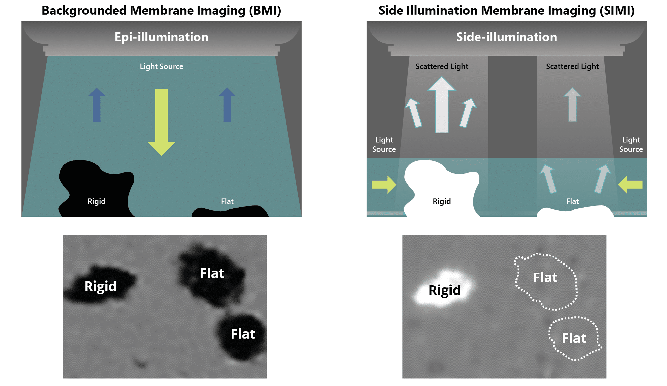
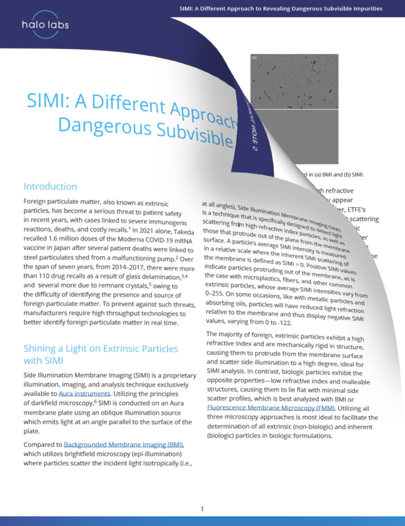
Side Illumination Membrane Imaging (SIMI) is a cutting-edge technology that allows for the detection of light scattering, specifically high refractive index particles, including those protruding out of the membrane surface.
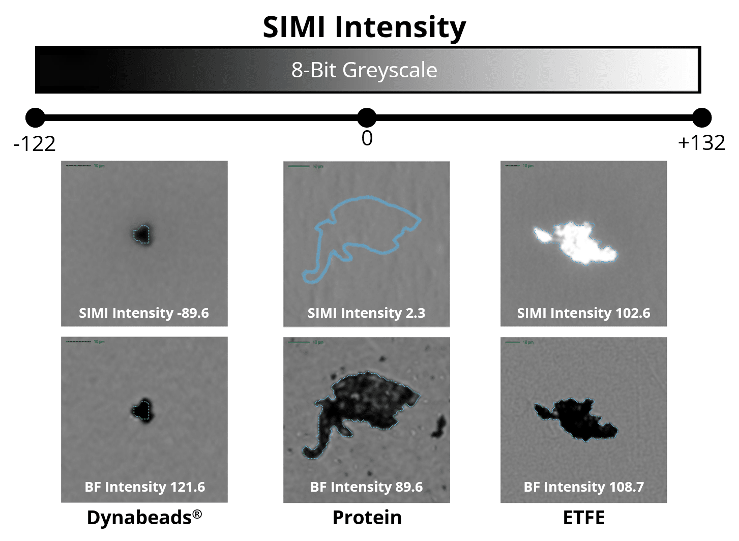
SIMI measures a particle's average intensity based on the amount of light that is scattered or absorbed from side illumination.
Particles that scatter light indicate inorganic matter protruding out of the membrane such as glass or plastic. Whereas particles that absorb light are indicative of metallic particles, such as residual Dynabeads™️, and absorbing oils yet also protrude from the membrane.
Foreign particles have high refractive index and rigid structures, making them ideal for SIMI analysis. However, biologic particles (e.g., proteins and viral vectors) have low refractive index and malleable structures, requiring Background Membrane Imaging (BMI) or Fluorescence Membrane Microscopy (FMM) analysis due to their minimal side scatter signature.
*Dynabeads refers to the magnetic beads produced by Thermo Fisher Scientific, Inc. Halo Labs is not affiliated with Thermo Fisher Scientific, Inc., and references to Dynabeads or any other third-party trademark do not imply sponsorship, endorsement, or approval.
Differentiate Organic and Inorganic Particles
SIMI can distinguish between organic and inorganic particles, making it ideal for interrogating heterogeneous particle solutions.
As a tertiary and complementary imaging mode to BMI and FMM, SIMI offers selective, orthogonal morphological data that can identify tall and rigid materials, which are defining features of most extrinsic particles.
While BMI is used for particle sizing and counting, SIMI provides the necessary illumination, imaging, and analysis to identify subvisible foreign particles across different biological products and bioprocess stages.
How SIMI Works With BMI
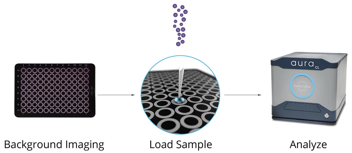
Particle analysis has never been simpler. With this innovative technology, you can screen up to 96 samples quickly and easily in just three steps:
- Backgrounded Membrane Imaging begins with imaging a new membrane plate.
- Pipetted samples are then placed into separate wells on the membrane, and a gentle vacuum is applied.
- Finally, load the plate once more and hit the "measure" button—it’s that straightforward.
- SIMI is automatically included with each BMI measurement, allowing you to characterize every particle in your sample each time you measure.


