Know What’s In Your Protein, Cell and Gene Therapies
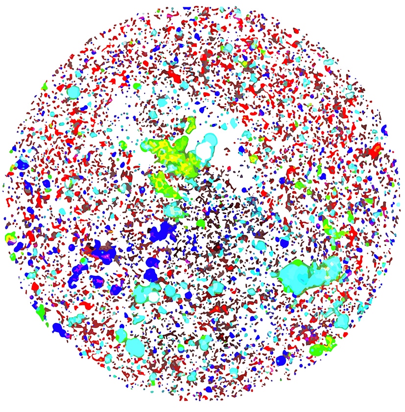
Characterizing biopharmaceutical products is no easy task, and traditional "counting techniques" just won't cut it. Regulatory agencies are calling for a more holistic approach—that's now possible with FMM, a particle-identifying technology exclusively available on Aura.
Now you can get 100% sampling efficiency at unparalleled speeds, making it possible to characterize and differentiate anything from a single aggregate protein or cell to tens of millions of particles in under two hours.
How FMM Works
The powerful combination of Fluorescence Membrane Microscopy (FMM) and Backgrounded Membrane Imaging (BMI) delivers an unmatched level of analysis to identify, count, and size particles that was never before possible.
In FMM, aggregates are labeled using specific fluorescent dyes or antibodies using flexible protocols that easily fit into your workflow, resulting in simple and definitive identification.
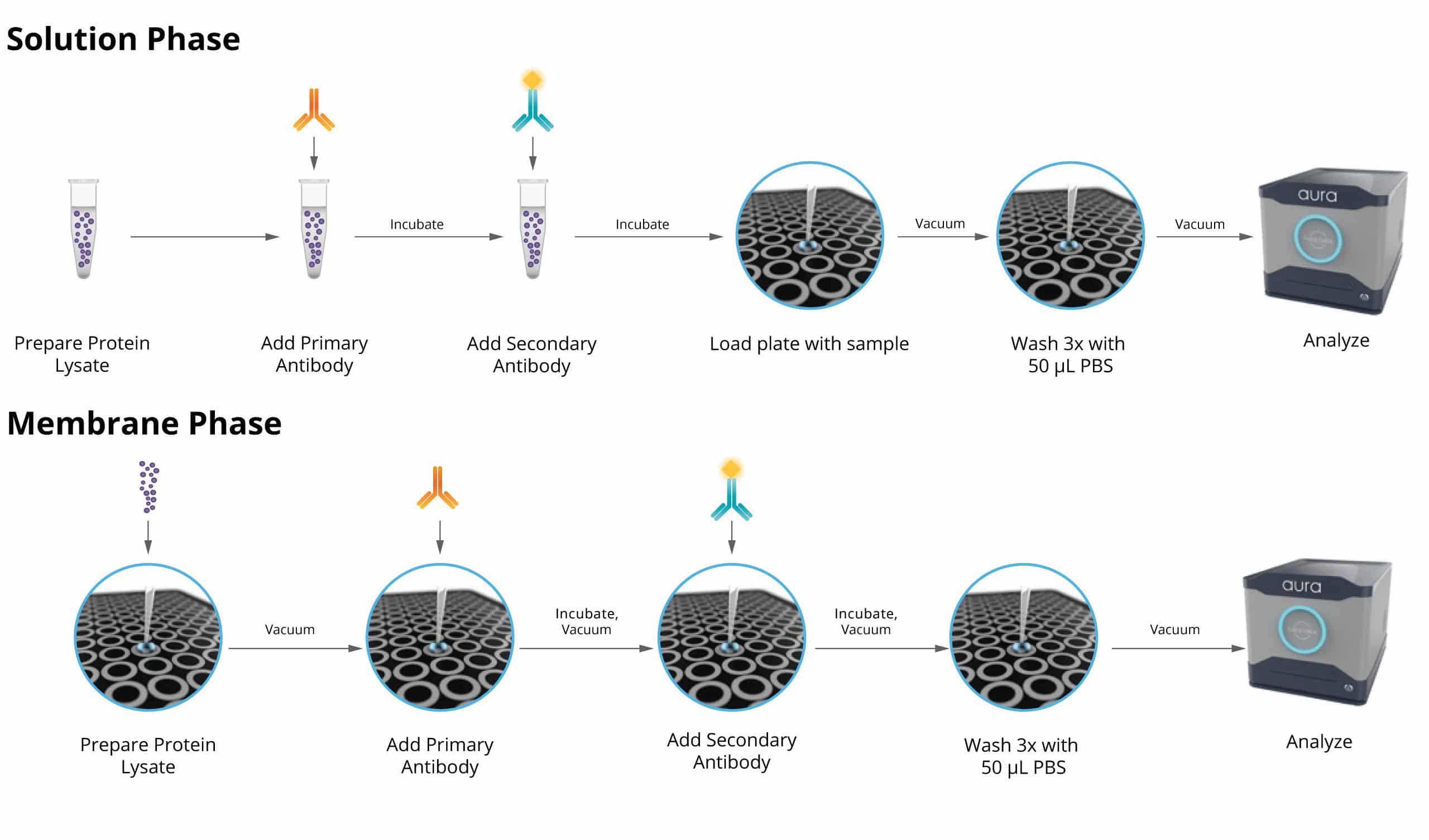
FMM Workflows
Solution Phase Stain
A fluorescent dye reagent is mixed with the sample in a test tube prior to applying it to a backgrounded plate. BMI and FMM measurements can then be taken.
Membrane Phase Stain
Microscopic particles are captured on the membrane surface and labeled with a fluorescent reagent. This can be accomplished using normal BMI steps with 40 µL of fluorescent reagent added to the sample with a second vacuum applied so the FMM measurement can be taken.


