Backgrounded Membrane Imaging: A Contender for Biologic USP 788 Subvisible Particle Analysis?
September 27, 2023
Biotherapeutic development heavily relies on the identification of visible and sub-visible particles to ensure product safety and efficacy. While conventional methods like Micro-Flow Imaging (MFI™) and Light Obscuration (LO) have been the industry standard for USP 788 particle analysis, a newcomer has stolen the spotlight, challenging these established norms: Backgrounded Membrane Imaging (BMI).
The Power of Background Membrane Imaging Rooted in USP 788 Principles
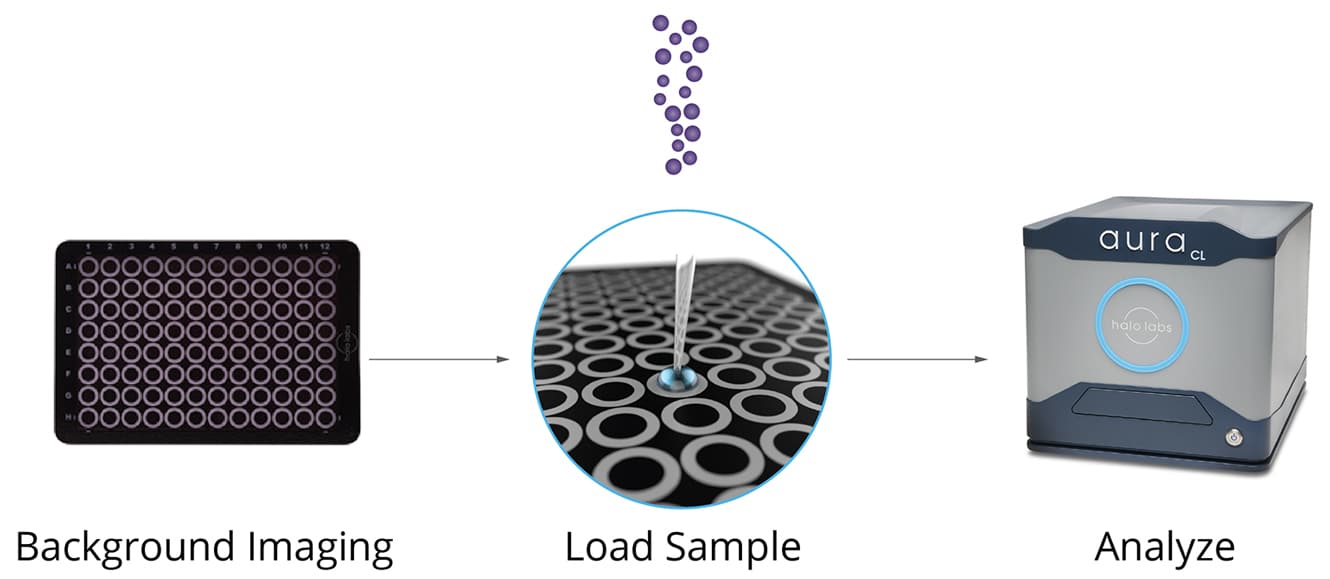
Leveraging the principles of USP 788 compendial method, Background Membrane Imaging diverges from traditional methods by using a membrane instead of a flow cell. This modernized approach offers a more accurate and cost-effective solution for particle identification and characterization. As a high-throughput microscopic technique, BMI can detect particles as small as 2 μm with just 25 μl of sample volume, making it an ideal choice when sample availability is limited.1
In the recent study “Backgrounded Membrane Imaging—A Valuable Alternative for Particle Detection of Biotherapeutics?” by Schleinzer et al., researchers compared BMI to MFI and LO methods.
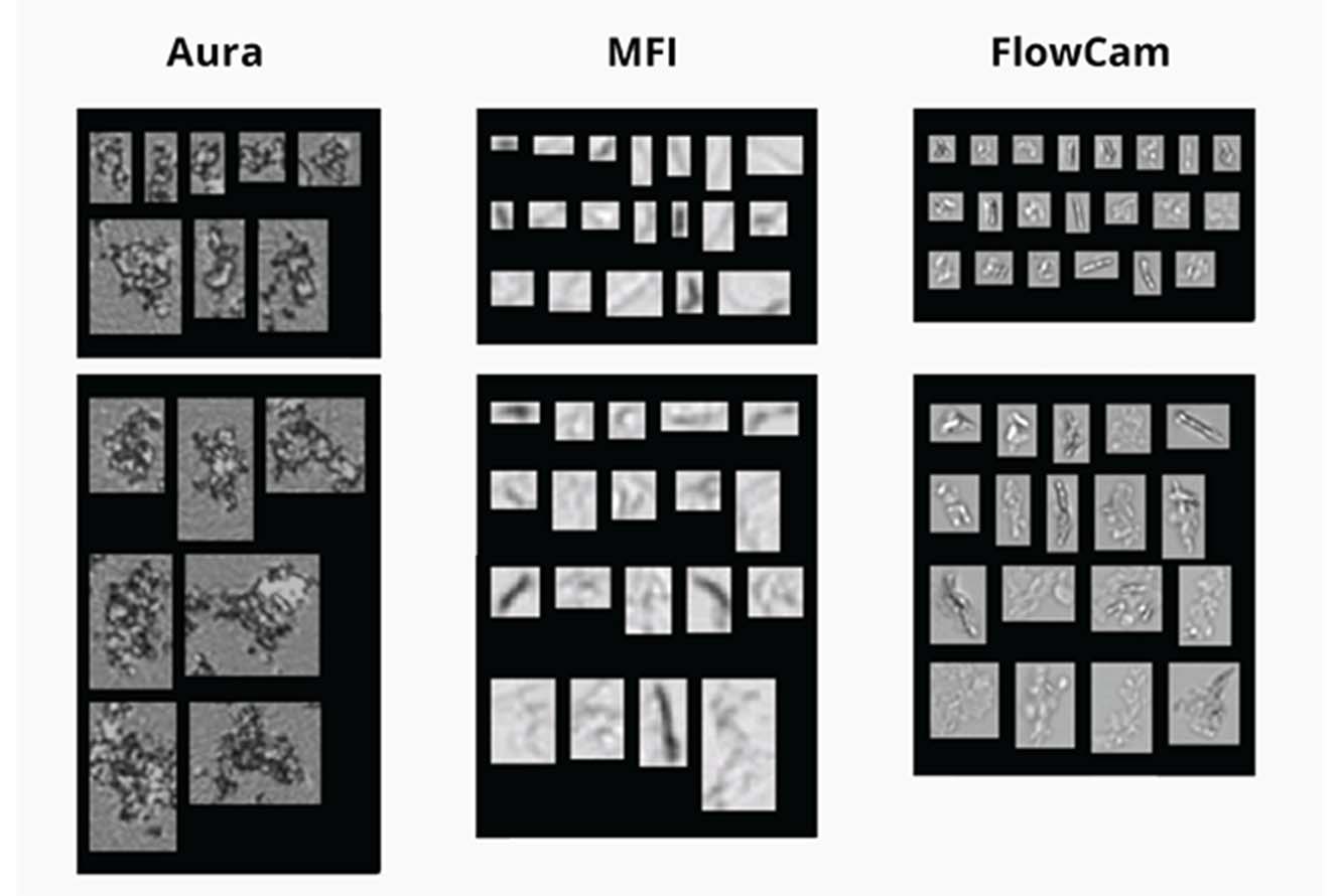
When analyzing stressed monoclonal antibody (mAb) samples, Background Membrane Imaging detected particle concentrations that were significantly higher (up to one to two orders of magnitude) than the measurements from MFI and light obscuration. This highlights the superior performance of Background Membrane Imaging in accurately detecting particle size, distribution, and concentration in biologics.1
Beyond its precision, Background Membrane Imaging stands out for its efficiency, measuring up to 96 samples in a single run. Preparing and measuring a 96-well plate takes approximately 3 hours, and no specific sample preparation prior to the experiment is required, further streamlining the process.
To analyze protein probes, a stressed mAb sample was examined under various formulation conditions using three techniques: BMI, MFI, and light obscuration. In these studies, BMI outperformed both MFI and light obscuration by detecting a significantly higher number of particles, exceeding them by one and two orders of magnitude for particles ≥ 2 μm. The same trend was observed for particles ≥ 10 μm and ≥ 25 μm. These findings highlight the superior sensitivity of BMI in detecting particles across different formulation conditions, comparable to MFI and light obscuration.1
Refractive Index Imaging: A Game-Changer in High Contrast Imaging
One of the key factors that contribute to the detection capabilities of Backgrounded Membrane Imaging is its manipulation of refractive index (ΔRI). By eliminating the formulation liquid phase, BMI enhances the difference in refractive index, resulting in particles that are less translucent and more easily detectable. This innovative technique allows for measurements with a significantly larger ΔRI of particles on the membrane than particles measured in the formulation buffer. The increased sensitivity of BMI for particle detection, especially for translucent particles, has already been well-established in various studies.1
The primary benefit of Backgrounded Membrane Imaging lies in its ability to enhance the visualization and detection of translucent protein particles by removing the liquid phase during sample filtration on the membrane. This leads to an increased refractive index difference (ΔRI), which enhances the contrast of each particle in the microscope image. Additionally, BMI eliminates air bubbles as a potential source of false positive particles, setting it apart from flow-based particle detection methods. Comprehensive studies have demonstrated a remarkable one order of magnitude increase in particle detection when using BMI compared to traditional MFI, and an impressive two orders of magnitude increase compared to LO.1
Advancing Subvisible Particle Analysis for Biotherapeutic Developers
Subvisible particle analysis technology has historically been limited, offering only a glimpse into what’s actually present in formulations, leaving crucial questions unanswered. At Halo Labs, we use Backgrounded Membrane Imaging (BMI), an advanced high-contrast imaging technique, in our Aura® systems which serves as the solution to these challenges.
BMI boasts exceptional detection capabilities, efficiency, and adaptability, making it a highly valuable alternative for particle analysis in the field of biotherapeutics. As we delve deeper into the potential of this cutting-edge technology, it becomes evident that BMI has the power to transform particle detection methods, setting a new benchmark for quality control within biotherapeutics.
References
1. Schleinzer, F., Strebl, M., Blech, M. et al.Backgrounded Membrane Imaging—A Valuable Alternative for Particle Detection of Biotherapeutics?. J Pharm Innov (2023). https://doi.org/10.1007/s12247-023-09734-5
DISCOVER THE AURA FAMILY
Read More ON this topic


