Backgrounded Membrane Imaging: A Breakthrough Approach for Subvisible Particle Characterization
March 27, 2024
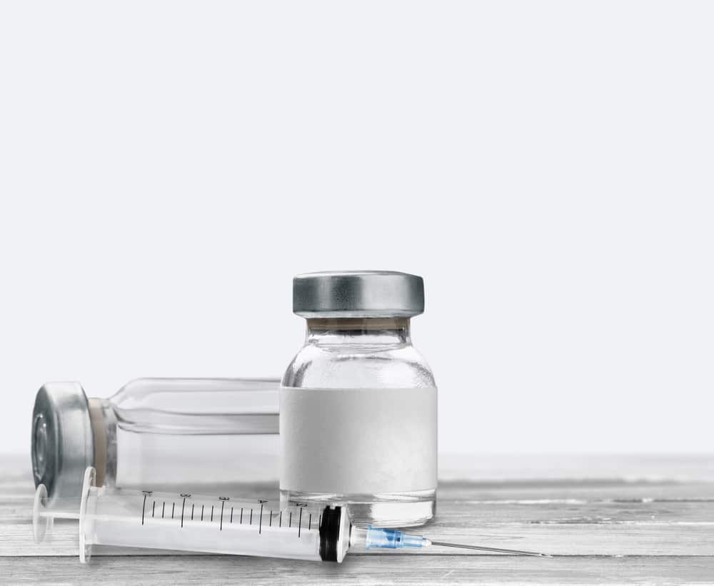
We know all too well how critical it is to address the potential harms that can arise with particulate matter in parenterals. It’s no secret that the presence of these particles—potentially resulting from protein instability, polysorbate degradation, or various other sources throughout manufacture, transport, storage, and administration—can hinder the efficacy and safety of drug products. 1
Light obscuration (LO) and the microscopic particle count test are the compendial methods for subvisible particle (SVP) quantification in parenteral drug products.1 The ICH-harmonized United States Pharmacopeia chapter <788> specifies limits for SVPs of sizes ≥10 μm and ≥25 μm. Moreover, regulatory authorities like the FDA expect the characterization of particles in the size range between 2 μm and 10 μm. In addition to the LO method, dynamic image analysis (DIA) is frequently applied to evaluate particulate matter in biopharma products.1
The challenge is that these techniques fail to adequately detect and measure SVPs. LO and DIA have limitations such as decreased sensitivity with low differences in refractive index and limited sample throughput. 1
Membrane microscopy can provide more reliable detection of SVPs exhibiting a refractive index close to their surrounding medium, and interferences by air bubbles or silicone oil droplets are not expected. However, this approach is labor intensive, and particle analysis is based on visual assessment and manual sizing by trained staff. 1
Evaluating the Efficacy of Backgrounded Membrane Imaging
In a recent study published in the Journal of Pharmaceutical Sciences, Backgrounded Membrane Imaging was evaluated in terms of its sizing and counting accuracy, working range, impact of refractive index, and interference caused by silicone oil droplets. This research also compared BMI to dynamic image analysis (DIA). Halo Labs’ Aura system was used to perform BMI. 1
Image quality was found to be comparable to current DIA methodologies. However, with the first versions of BMI image analysis software, an under sizing of polystyrene beads was observed. Please note several new algorithms and software versions have been released addressing this issue (see Tech Note 3 for more information). BMI linear concentration range was found to reach an upper limit (7.1 x 10⁵ particles/mL) similar to DIA. In the absence of silicone oil droplets, BMI and DIA showed good agreement in total particle concentrations (particle diameter ≥2 mm) but differences in size distributions for particle sizes ≥4 mm. 1
Analyses of prefilled syringe products and silicone oil emulsions demonstrated the removal of silicone oil in BMI sample processing. In contrast to DIA, particle counting by BMI remained unaffected by changes in refractive index. 1
Overall, BMI demonstrated to be a promising orthogonal method for subvisible particle characterization. Aspects like low required sample volume, high throughput, and ease of handling can make BMI a valuable alternative or complement to DIA in particular for formulation screening. 1
The study also pointed out that BMI can be a useful tool for high-throughput screenings, such as in formulation development where different formulations need to be compared. The removal of silicone oil droplets during BMI sample preparation is beneficial for the analysis of drug products from prefilled syringes as it ensures only non-silicone oil particles are counted. The authors recommended adaptations to the BMI system to enable full well imaging for accurate particle counting and improvements in the BMI/Aura software for better particle recognition and reduced fragmentation. 1
The Promise of Backgrounded Membrane Imaging
Detecting and quantifying subvisible particles can be quite challenging when the refractive index is so close to its surrounding medium. To tackle this issue, Halo Labs has developed Backgrounded Membrane Imaging (BMI), a high-contrast imaging technique. As a contemporary form of membrane microscopy, BMI offers an automated, 96-well plate-based approach to microscopic particle analysis (in the size range starting from ~2 mm).
BMI utilizes sophisticated image processing techniques to analyze images and acquire particle data. Its working principle is based on acquiring images of custom-designed polycarbonate membrane filter plates before and after sample application. This creates a background image and an actual measurement image, which are aligned and subtracted from each other by image analysis software. The result is an isolated image of the actual particles, ready for analysis. 1
Liquid samples (~20–150 mL) are pipetted onto the membrane wells defined by hydrophobic rings. By the application of vacuum suction, liquid is drawn through the membrane pores and removed from the samples, leaving particles on the membrane surface for subsequent image analysis. 1
Conclusion
It’s evident that backgrounded membrane imaging is a promising orthogonal technique to dynamically detect and characterize potentially harmful subvisible particulates in biopharmaceuticals. 1 With its simplicity, high throughput, and sensitivity, BMI enables accelerated biopharmaceutical research with enhanced particulate monitoring—making drug discovery and development smarter and safer.
Related
- Join our virtual course and gain the skills to load samples, image, count, and size subvisible particles using Backgrounded Membrane Imaging
- See how BMI provides valuable insights into particle analysis
- Are you missing the complete picture by using light obscuration?
References
- Helbig, C., Ammann, G., Menzen, T., Friess, W., Wuchner, K., & Hawe, A. (2020). Backgrounded Membrane Imaging (BMI) for High-Throughput Characterization of Subvisible Particles During Biopharmaceutical Drug Product Development. Journal of Pharmaceutical Sciences, 109(1), 264-276. https://doi.org/10.1016/j.xphs.2019.03.024
")