Virtual Seminar – AAV Vector Stability With BMI – Are Your Analytics Giving You The Whole Picture? – April 2020
Subvisible particle (SVP) analysis in parenteral formulations is a key predictor of safety and efficacy, and an essential drug quality metric. For viral vectors, however, limited availability of sample makes measuring SVP’s a challenge. Standard methods such as light obscuration and flow imaging are impractical due to their large volume requirements and low throughput.
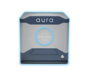
This seminar will introduce automated Backgrounded Membrane Imaging (BMI) as a solution for detecting and quantifying subvisible AAV aggregates in low volume, high throughput format. BMI is fully automated, fluidics-free, and uses only 25 μL per sample. Learn how BMI’s high-resolution images and comprehensive data analysis can reveal particle issues that may be missed by other methods like DLS and SEC – providing a more complete picture of your viral vector stability


