The First Technology to Accurately Detect Residual Dynabeads in Cell Therapies
CAR-T cell manufacturing often requires the use of Dynabeads™️ for selection and activation, but their residual presence in a finished drug product can put patient safety at risk.
Without reliable methods to detect remaining Dynabeads, manual hemocytometry processes are often inadequate and subjective, introducing errors and bead undercounting into an already complex process.
With Aura CL and Aura+, we're empowering cell therapy developers to make safer drugs with the industry's first technology that identifies residual Dynabeads in highly concentrated products.
*Dynabeads refers to the magnetic beads produced by Thermo Fisher Scientific, Inc. Halo Labs is not affiliated with Thermo Fisher Scientific, Inc., and references to Dynabeads or any other third-party trademark do not imply sponsorship, endorsement, or approval.

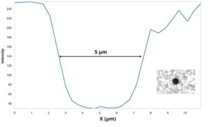
Dynabeads size: 5 µm
Why Use Aura to Detect and Count Residual Dynabeads?
- Best in class Dynabeads identification and quantitation
- Accurate, fluidics-free cell analysis at high concentrations
- Obtain detailed information on particles that other methods can’t deliver, including size, morphology, count, and distribution
- Rapid analysis time of about 1 minute per sample
- Particles are imaged without the interference of buffer or matrix for higher sensitivity
- Achieve high sensitivity because particles are imaged without the interference of buffer or matrix
- Automated data analysis
- USP 788 Method 2 and USP 1046 compliant
- Maintain compliance with the option for 21 CFR Part 11 software
Quickly Distinguish Dynabeads from Other Particles
Aura CL provides a comprehensive characterization of all particles in cell therapy products, powered by innovative Backgrounded Membrane Microscopy (BMI) and Side Illuminated Membrane Imaging (SIMI) technologies to detect contaminants like Dynabeads that have persisted throughout the manufacturing process.
With BMI and SIMI working together, you get a fuller picture of highly concentrated cells with single digit counts of Dynabeads, respectively.
Count Dynabeads with Greater Accuracy and High Throughput
With Aura CL, users have the power to make smart decisions within a crowded cell space.
Unlike other methods that are inefficient and prone to errors, this tool offers greater accuracy and higher-throughput counting capabilities.
And SIMI takes automation one step further with its 'x-ray' vision—allowing it to identify even a single Dynabeads among hundreds of thousands of cells.

Particle Vue Software
Get particle analysis answers in just a few clicks with flexible, easy-to-use Particle Vue Software.
Featured products

Aura+
With the ability to support the full range of development and manufacturing applications across therapeutic approaches—including protein/antibody therapeutics, gene therapies, cell therapies, and small molecules— Aura+ packs powerful flexibility into a single particle analyzer.
Features
- 5 µL to 10 mL volume
- Particle characterization for protein, cell and gene therapeutics
- Three fluorescent channels for particle identification
- 4x and 20x magnification imaging

Aura CL
Safeguard your cell therapies from contamination with Aura CL. Our high-throughput, fluidic-free particle analysis system quickly and accurately identifies and characterizes subvisible particles, giving you peace of mind when it comes to cell therapy development and quality assessment.
Features
- Automated residual Dynabeads analysis
- Cell purity and viability characterization
- High powered magnification
- Fluidics free
Frequently Asked Questions
Dynabeads are small, spherical beads that have been widely used in scientific research and medical applications. Developed by Thermo Fisher Scientific, Dynabeads are composed of a magnetic core and a surface coated with a matrix that specifically binds to various substances, such as proteins, antibodies, or nucleic acids. Due to their unique magnetic properties, Dynabeads can be easily manipulated using a magnetic field. These beads have revolutionized numerous biological techniques by enabling researchers to efficiently isolate, separate, and manipulate specific target molecules from complex mixtures. Their diverse range of applications includes biomarker discovery, immunoprecipitation, cell isolation, and gene expression analysis. Overall, Dynabeads have become invaluable tools in the field of life sciences, enabling researchers to gain deeper insights and advance various areas of study by simplifying and enhancing experimental processes.
Dynabeads typically come in 2.8, 4.5 or 1.0 µm sizes. Most of our customers use 4.5µm beads often referring to them as "5µm beads."
Typical 5µm Dynabeads sized beads measure as SIMI negative, 5-pixel profiles. For more information, download Tech Note 4.
Aura can detect any magnetic bead or Dynabeads size 1.0 µm or greater. In the case of Thermofisher Dynabeads offerings, Aura can detect, ID, count and size all of their magnetic bead products.
Generically speaking, Aura can detect and ID any magnetic beads similar to Dynabeads. Aura can also go beyond magnetics beads, capable of looking for other beads such as polystyrene and even microcarriers.


")